


- Who we serve
- Academia
Explore BioLegend
Learn about our world class antibodies for a diverse set of research areas including immunology, neuroscience, cancer, stem cells and cell biology.


- Pharma / Biotech
- Clinical Laboratories
- Healthcare Professionals
- Contract Research Organizations
- Academia
- Products
- Research & Development
- Clinical & Diagnostics
- Reproductive Health
- Infectious Diseases
- Cancer
- Autoimmunity
- Endocrinology
- Allergy
- Neurodegeneration
- Rapid Patient Testing
- Reproductive Health
- Reagents
- Platforms & Automation
- Nucleic Acid Purification
- Automated Liquid Handling
- Integrated Lab Automation
- Microfluidic Analysis
- Detection Solutions
- Imaging
- Sample Homogenization
- In Vitro Diagnostics (IVD) Platforms & Automation
- Consumables & Accessories
- Instrument Service & Maintenance
- Nucleic Acid Purification
- Consumables & Accessories
- Signals Software
- Revvity Omics Services
- Research & Development
- Services
- Preclinical Services
- Antibody Drug Conjugate Services
Preclinical services
Work with our experienced scientific team and leverage our advanced technologies to help accelerate the preclinical drug discovery process.

- Complex Cell Model Screening Services
Preclinical services
Work with our experienced scientific team and leverage our advanced technologies to help accelerate the preclinical drug discovery process.
- Base Editing Platform
Preclinical services
Work with our experienced scientific team and leverage our advanced technologies to help accelerate the preclinical drug discovery process.
- Immune Cell Screening
Preclinical services
Work with our experienced scientific team and leverage our advanced technologies to help accelerate the preclinical drug discovery process.
- Functional Genomic Screening Services
Preclinical services
Work with our experienced scientific team and leverage our advanced technologies to help accelerate the preclinical drug discovery process.
- Cell Panel Screening
Preclinical services
Work with our experienced scientific team and leverage our advanced technologies to help accelerate the preclinical drug discovery process.
- Cell Line Engineering
Preclinical services
Work with our experienced scientific team and leverage our advanced technologies to help accelerate the preclinical drug discovery process.
- Viral Vector Engineering and Manufacture
Preclinical services
Work with our experienced scientific team and leverage our advanced technologies to help accelerate the preclinical drug discovery process.
Preclinical services
Work with our experienced scientific team and leverage our advanced technologies to help accelerate the preclinical drug discovery process.
- Antibody Drug Conjugate Services
- Revvity Omics Services
- Revvity Omics Clinical Services
T-SPOT.TB testing services.
Revvity's Oxford Diagnostic Laboratories is a large referral laboratory for tuberculosis testing services based on our T-SPOT technology.

- Revvity Omics Pharma Services
T-SPOT.TB testing services.
Revvity's Oxford Diagnostic Laboratories is a large referral laboratory for tuberculosis testing services based on our T-SPOT technology.
T-SPOT.TB testing services.
Revvity's Oxford Diagnostic Laboratories is a large referral laboratory for tuberculosis testing services based on our T-SPOT technology.
- Revvity Omics Clinical Services
- Clinical & Testing Services
- Revvity Omics Clinical Services
T-SPOT.TB testing services.
Revvity's Oxford Diagnostic Laboratories is a large referral laboratory for tuberculosis testing services based on our T-SPOT technology.
- Revvity Omics Pharma Services
T-SPOT.TB testing services.
Revvity's Oxford Diagnostic Laboratories is a large referral laboratory for tuberculosis testing services based on our T-SPOT technology.
- Cellular and Humoral Immunoassays
T-SPOT.TB testing services.
Revvity's Oxford Diagnostic Laboratories is a large referral laboratory for tuberculosis testing services based on our T-SPOT technology.
- Tuberculosis Testing Services
T-SPOT.TB testing services.
Revvity's Oxford Diagnostic Laboratories is a large referral laboratory for tuberculosis testing services based on our T-SPOT technology.
T-SPOT.TB testing services.
Revvity's Oxford Diagnostic Laboratories is a large referral laboratory for tuberculosis testing services based on our T-SPOT technology.
- Revvity Omics Clinical Services
- Customization Services
- Assays and Reagents
T-SPOT.TB testing services.
Revvity's Oxford Diagnostic Laboratories is a large referral laboratory for tuberculosis testing services based on our T-SPOT technology.
- Microplate Services
T-SPOT.TB testing services.
Revvity's Oxford Diagnostic Laboratories is a large referral laboratory for tuberculosis testing services based on our T-SPOT technology.
- Custom Conjugation & Labeling
T-SPOT.TB testing services.
Revvity's Oxford Diagnostic Laboratories is a large referral laboratory for tuberculosis testing services based on our T-SPOT technology.
- Radiosynthesis and Labeling
T-SPOT.TB testing services.
Revvity's Oxford Diagnostic Laboratories is a large referral laboratory for tuberculosis testing services based on our T-SPOT technology.
T-SPOT.TB testing services.
Revvity's Oxford Diagnostic Laboratories is a large referral laboratory for tuberculosis testing services based on our T-SPOT technology.
- Assays and Reagents
- Licensing
- Gene Delivery Licensing
CHOSOURCE expression platform
Revvity's expression platform provides an enhanced system for the development and manufacturing of biotherapeutics that can be used in commercial manufacturing applications.

- Gene Expression Systems
CHOSOURCE expression platform
Revvity's expression platform provides an enhanced system for the development and manufacturing of biotherapeutics that can be used in commercial manufacturing applications.
- Pin-point Base Editing Platform
CHOSOURCE expression platform
Revvity's expression platform provides an enhanced system for the development and manufacturing of biotherapeutics that can be used in commercial manufacturing applications.
- Virus Screening
CHOSOURCE expression platform
Revvity's expression platform provides an enhanced system for the development and manufacturing of biotherapeutics that can be used in commercial manufacturing applications.
CHOSOURCE expression platform
Revvity's expression platform provides an enhanced system for the development and manufacturing of biotherapeutics that can be used in commercial manufacturing applications.
- Gene Delivery Licensing
- Viral Vector Engineering and Manufacture
- AAV Services
T-SPOT.TB testing services.
Revvity's Oxford Diagnostic Laboratories is a large referral laboratory for tuberculosis testing services based on our T-SPOT technology.
- Lentivirus Services
T-SPOT.TB testing services.
Revvity's Oxford Diagnostic Laboratories is a large referral laboratory for tuberculosis testing services based on our T-SPOT technology.
T-SPOT.TB testing services.
Revvity's Oxford Diagnostic Laboratories is a large referral laboratory for tuberculosis testing services based on our T-SPOT technology.
- AAV Services
- Instrument Service & Maintenance
- AV Services
T-SPOT.TB testing services.
Revvity's Oxford Diagnostic Laboratories is a large referral laboratory for tuberculosis testing services based on our T-SPOT technology.
- Equipment Service Plans
T-SPOT.TB testing services.
Revvity's Oxford Diagnostic Laboratories is a large referral laboratory for tuberculosis testing services based on our T-SPOT technology.
- On-demand Equipment Service
T-SPOT.TB testing services.
Revvity's Oxford Diagnostic Laboratories is a large referral laboratory for tuberculosis testing services based on our T-SPOT technology.
T-SPOT.TB testing services.
Revvity's Oxford Diagnostic Laboratories is a large referral laboratory for tuberculosis testing services based on our T-SPOT technology.
- AV Services
- Customer Training
- Expert-led Training
T-SPOT.TB testing services.
Revvity's Oxford Diagnostic Laboratories is a large referral laboratory for tuberculosis testing services based on our T-SPOT technology.
- Online Training
T-SPOT.TB testing services.
Revvity's Oxford Diagnostic Laboratories is a large referral laboratory for tuberculosis testing services based on our T-SPOT technology.
T-SPOT.TB testing services.
Revvity's Oxford Diagnostic Laboratories is a large referral laboratory for tuberculosis testing services based on our T-SPOT technology.
- Expert-led Training
- OEM Solutions
T-SPOT.TB testing services.
Revvity's Oxford Diagnostic Laboratories is a large referral laboratory for tuberculosis testing services based on our T-SPOT technology.
T-SPOT.TB testing services.
Revvity's Oxford Diagnostic Laboratories is a large referral laboratory for tuberculosis testing services based on our T-SPOT technology.
- Preclinical Services
- Company
- Purpose
- Careers
- Investor Relations
- Events
Investor Day 2024
Driving meaningful innovation that profoundly impacts science and human lives.

- Financials
Investor Day 2024
Driving meaningful innovation that profoundly impacts science and human lives.
- Stock Info
Investor Day 2024
Driving meaningful innovation that profoundly impacts science and human lives.
Investor Day 2024
Driving meaningful innovation that profoundly impacts science and human lives.
- Events
- ESG
- News
- Purpose
- Resources
- Product Support
- Application Support Knowledge base (ASK)
Tech documents, at your fingertips.
Quickly find and download manuals, safety documents, certificates of analysis and more.

- SDS Search
Tech documents, at your fingertips.
Quickly find and download manuals, safety documents, certificates of analysis and more.
- COA/TDS Search
Tech documents, at your fingertips.
Quickly find and download manuals, safety documents, certificates of analysis and more.
- Manual/IFU Search
Tech documents, at your fingertips.
Quickly find and download manuals, safety documents, certificates of analysis and more.
- SpectraViewer
Tech documents, at your fingertips.
Quickly find and download manuals, safety documents, certificates of analysis and more.
- RAD Calculator
Tech documents, at your fingertips.
Quickly find and download manuals, safety documents, certificates of analysis and more.
Tech documents, at your fingertips.
Quickly find and download manuals, safety documents, certificates of analysis and more.
- Application Support Knowledge base (ASK)
- Resource Center
Tech documents, at your fingertips.
Quickly find and download manuals, safety documents, certificates of analysis and more.
- Blog
Tech documents, at your fingertips.
Quickly find and download manuals, safety documents, certificates of analysis and more.
- Events
Tech documents, at your fingertips.
Quickly find and download manuals, safety documents, certificates of analysis and more.
- Customer Training
- Expert-led Training
Tech documents, at your fingertips.
Quickly find and download manuals, safety documents, certificates of analysis and more.
- Online Training
Tech documents, at your fingertips.
Quickly find and download manuals, safety documents, certificates of analysis and more.
Tech documents, at your fingertips.
Quickly find and download manuals, safety documents, certificates of analysis and more.
- Expert-led Training
- Help Center
- Order Support
Tech documents, at your fingertips.
Quickly find and download manuals, safety documents, certificates of analysis and more.
- Contact Us
Tech documents, at your fingertips.
Quickly find and download manuals, safety documents, certificates of analysis and more.
- Technical Support
Tech documents, at your fingertips.
Quickly find and download manuals, safety documents, certificates of analysis and more.
- Instruments Support & Service
Tech documents, at your fingertips.
Quickly find and download manuals, safety documents, certificates of analysis and more.
- SDS Request
Tech documents, at your fingertips.
Quickly find and download manuals, safety documents, certificates of analysis and more.
- COA/TDS Request
Tech documents, at your fingertips.
Quickly find and download manuals, safety documents, certificates of analysis and more.
- Manual/IFU Request
Tech documents, at your fingertips.
Quickly find and download manuals, safety documents, certificates of analysis and more.
- Training Request
Tech documents, at your fingertips.
Quickly find and download manuals, safety documents, certificates of analysis and more.
- Cell Line Terms & Conditions
Tech documents, at your fingertips.
Quickly find and download manuals, safety documents, certificates of analysis and more.
Tech documents, at your fingertips.
Quickly find and download manuals, safety documents, certificates of analysis and more.
- Order Support
- FAQs
Tech documents, at your fingertips.
Quickly find and download manuals, safety documents, certificates of analysis and more.
- Software Downloads
Tech documents, at your fingertips.
Quickly find and download manuals, safety documents, certificates of analysis and more.
- Knowledge Base
- Application support knowledge base (ASK)
Tech documents, at your fingertips.
Quickly find and download manuals, safety documents, certificates of analysis and more.
- Newborn screening disorders
Tech documents, at your fingertips.
Quickly find and download manuals, safety documents, certificates of analysis and more.
- Sample homogenization applications and protocols
Tech documents, at your fingertips.
Quickly find and download manuals, safety documents, certificates of analysis and more.
- TB testing services
Tech documents, at your fingertips.
Quickly find and download manuals, safety documents, certificates of analysis and more.
Tech documents, at your fingertips.
Quickly find and download manuals, safety documents, certificates of analysis and more.
- Application support knowledge base (ASK)
Tech documents, at your fingertips.
Quickly find and download manuals, safety documents, certificates of analysis and more.
- Product Support
Welcome to Revvity: renowned brands and boundless innovation.
Hearing the word "can't" is our call to action!
We help scientists, researchers, and clinicians overcome the world's greatest health obstacles.
View our story
Featured brand: BioLegend
Learn about our world-class antibodies for a diverse set of research areas including immunology, neuroscience, cancer, stem cells and cell biology.
Visit BioLegend.com

US
Revvity Sites Globally
Select your location.
*e-commerce not available for this region.

HTRF Human Phospho-BAD (Ser112) Detection Kit, 500 Assay Points


HTRF Human Phospho-BAD (Ser112) Detection Kit, 500 Assay Points


Shipping box for Revvity reagent kits


The Phospho-BAD (Ser112) kit enables the cell-based quantification of phosphorylated BAD (Ser112) as a marker of cells entering apoptosis.
| Feature | Specification |
|---|---|
| Application | Cell Signaling |
| Sample Volume | 16 µL |
The Phospho-BAD (Ser112) kit enables the cell-based quantification of phosphorylated BAD (Ser112) as a marker of cells entering apoptosis.
Product Variants
Unit Size: 96 assay points
Part #:
64BADPET
List Price
USD 703.20
Your online price:
Unit Size: 500 assay points
Part #:
64BADPEG
List Price
USD 2,271.53
Your online price:
Unit Size: 10,000 assay points
Part #:
64BADPEH
List Price
USD 13,214.42
Your online price:
For research use only. Not for use in diagnostic procedures. All products to be used in accordance with applicable laws and regulations including without limitation, consumption, and disposal requirements under European REACH regulations (EC 1907/2006).
HTRF Human Phospho-BAD (Ser112) Detection Kit, 500 Assay Points
Shipping box for Revvity reagent kits
HTRF Human Phospho-BAD (Ser112) Detection Kit, 500 Assay Points
Product information
Overview
The Phospho-Bad (Ser112) cellular kit is designed for the streamlined and efficient quantification of phosphorylated BAD proteins in cell lysates.This fast and convenient, no-wash protocol offers highly sensitive results and can be applied to all steps of the drug discovery process, from basic research to High Throughput screening.
Specifications
| Application |
Cell Signaling
|
|---|---|
| Brand |
HTRF
|
| Detection Modality |
HTRF
|
| Lysis Buffer Compatibility |
Lysis Buffer 3
|
| Molecular Modification |
Phosphorylation
|
| Product Group |
Kit
|
| Sample Volume |
16 µL
|
| Shipping Conditions |
Shipped in Dry Ice
|
| Target Class |
Phosphoproteins
|
| Target Species |
Human
|
| Technology |
TR-FRET
|
| Therapeutic Area |
Oncology & Inflammation
|
| Unit Size |
500 assay points
|
Video gallery
HTRF Human Phospho-BAD (Ser112) Detection Kit, 500 Assay Points
HTRF Human Phospho-BAD (Ser112) Detection Kit, 500 Assay Points
How it works
Phospho-BAD (Ser112) Assay principle
The Phospho-BAD (Ser112) assay measures BAD when phosphorylated at Ser112. Contrary to Western Blot, the assay is entirely plate-based and does not require gels, electrophoresis or transfer. The Phospho-BAD (Ser112) assay uses 2 labeled antibodies: one with a donor fluorophore, the other one with an acceptor. The first antibody is selected for its specific binding to the phosphorylated motif on the protein, the second for its ability to recognize the protein independent of its phosphorylation state. Protein phosphorylation enables an immune-complex formation involving both labeled antibodies and which brings the donor fluorophore into close proximity to the acceptor, thereby generating a FRET signal. Its intensity is directly proportional to the concentration of phosphorylated protein present in the sample, and provides a means of assessing the proteins phosphorylation state under a no-wash assay format.
Phospho-BAD (Ser112) 2-plate Assay protocol
The 2 plate protocol involves culturing cells in a 96-well plate before lysis then transferring lysates to a 384-well low volume detection plate before adding phospho-BAD (Ser112) HTRF detection reagents. This protocol enables the cells' viability and confluence to be monitored.

Phospho-BAD (Ser112) 1-plate assay protocol
Detection of Phosphorylated BAD (Ser112) with HTRF reagents can be performed in a single plate used for culturing, stimulation and lysis. No washing steps are required. This HTS designed protocol enables miniaturization while maintaining robust HTRF quality.

Assay validation
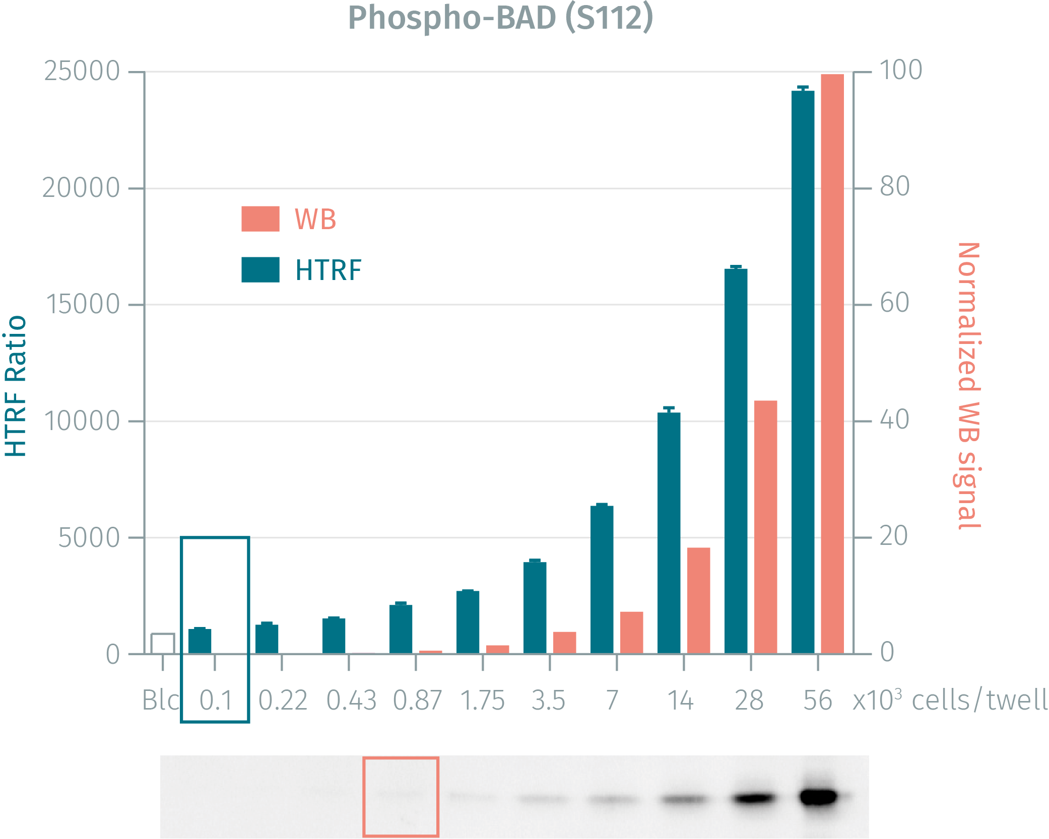
WB versus HTRF assay for Phoshpo-BAD (Ser112) kit
Cos-7 cells were grown in a T175 flask at 37°C, 5% CO2, for 1 day. Day 2: After removal of cell culture medium, 3 mL of supplemented lysis buffer were added and incubated for 30 minutess. Soluble supernatants were collected after a 10 minute centrifugation. Equal amounts of lysates were used for a side by side comparison of Western Blot and HTRF.220 cells can be detected by using HTRF phospho-Bad (Ser112) wheras1750 cells are needed for the Western Blot. The HTRF assay is 4-fold more sensitive than Western Blot.
PMA dose-response on HEK293 cells for Phospho-BAD (Ser112) kit
Human HEK293 cells (100,000 cells/well) were stimulated for 30 minutess at 37°C with various concentrations of PMA. After 30 minutess of lysis incubation, phosphorylated Bad was measured using the two-plate assay protocol.

Staurosporine inhibition on stimulated MCF7 and Cos-7 cells
Human MCF7 and monkey Cos-7 cells (25,000 cells/well) were incubated for 3 hours at 37°C with various concentrations of Staurosporin inhibitor. Then 0.8 µM of PMA was added for cell stimulation, and sampes were incubated for 30 minutess. After 30 minutess of lysis incubation, inhibition of Bad phosphorylation was measured using the HTRF Phospho-Bad (Ser112) assay with the two-plate protocol.

Simplified pathway
Simplified pathway of pro-apoptotic factor Bad signaling
Bad is a pro-apoptotic factor belonging to the Bcl-2 family of proteins and governs mitochondrial membrane permeability by regulating cytochrome C release. In healthy, proliferating cells, Bad is phosphorylated and sequestered in the cytosol while in stressed cells, death stimuli induce Bad dephosphorylation and its translocation to the mitochondrial membrane, where it neutralizes Bcl-Xl or BCl-2 by heterodimerization and thus induces cytochrome C release, leading to apoptosis.

Resources
Are you looking for resources, click on the resource type to explore further.
Brochure
HTRF assays and reagents catalog
Discover the versatility and precision of Homogeneous Time-Resolved Fluorescence (HTRF) technology. Our HTRF portfolio offers a...
Guide
HTRF solutions, guide to major applications
This guide provides you an overview of HTRF applications in several therapeutic areas.
SDS, COAs, Manuals and more
Are you looking for technical documents related to the product? We have categorized them in dedicated sections below. Explore now.
Safety data sheet
- LanguageEnglishCountryUnited States
- LanguageFrenchCountryFrance
- LanguageGermanCountryGermany
+ Show next 4
Certificate of analysis
- Lot Number06CLot DateDecember 14, 2025
- Lot Number06BLot DateNovember 9, 2024
- Lot Number06ALot DateNovember 9, 2024
+ Show next 2
IFUs, Manuals & more
- Resource TypeManualLanguageEnglishCountry-


Recently Viewed

USD 679.88 - USD 10,722.18
How can we help you?
We are here to answer your questions.

