


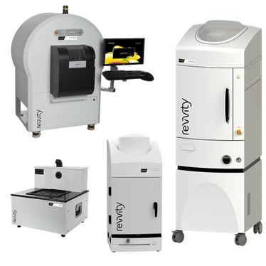

Vega preclinical ultrasound
- Hands-free, automated transducer positioning and movement
- High speed, high-throughput performance with 3 mice scanning in just a few minutes
- 3D Widefield acquisitions enabling whole subject imaging
- Standard B-mode and M-mode capability
- Shear Wave Elastography (SWE) mode for quantifying and evaluation tissue stiffness
- Acoustic angiography mode for visualizing microvasculature
- Fits on the benchtop

Vega Preclinical Ultrasound System
- Hands-free - Automated transducer positioning and movement
- Easy-to-use requiring minimal training
- High-speed, high-throughput performance with 3 mice scanning in just a few minutes
- 3D widefield acquisitions enabling whole subject imaging
- Standard B-Mode and M-Mode capability
- Shear Wave Elastography (SWE) mode for quantifying tissue stiffness
- Acoustic Angiography (AA) mode for visualization of microvasculature
- Flexible visualization and analysis software
- Fits on the benchtop
IVIS Lumina S5 & X5
- High-sensitivity 2D optical imaging (bioluminescence and fluorescence)
- High-throughput format with a 20 x 20 FOV sufficient for imaging 5 mice simultaneously
- High resolution, low dose X-ray with optical overlay (IVIS Lumina X5 only)
- Compact design that fits on your benchtop
- Unique animal handling accessories and software tools to streamline throughput
- Complimentary Living Image™ software licenses are provided with the IVIS systems and upon request.

IVIS Lumina S5 Imaging System
- High throughput (5 mice) optical imaging
- Increased throughput (10 mice) using optical manifold
- Supports mouse and rat imaging
- Compute Pure Spectrum (CPS) spectral unmixing
- Full fluorescence tunability through the NIR spectrum
- Unique accessories to speed acquisition and analysis
- Small footprint–sits on your benchtop
- Complimentary Living Image software licenses are provided with the IVIS systems and upon request.
IVIS Lumina X5 Imaging System
- High throughput (5 mice) optical and X-ray
- Increased throughput (10 mice) using optional manifold
- High resolution, low dose X-ray with optical overlay
- Supports mouse and rat imaging
- Compute Pure Spectrum (CPS) spectral unmixing
- Full fluorescence tunability through the NIR spectrum
- Unique accessories to streamline workflow, data acquisition and analysis
- Complimentary Living Image software licenses are provided with the IVIS systems and upon request
IVIS Lumina III series
- 2D optical imaging (bioluminescence and fluorescence)
- Low-dose X-ray with optical overlay (IVIS Lumina XRMS only)
- Images up to 3 mice simultaneously
- Compact design that fits on your benchtop
- Complimentary Living Image™ software licenses are provided with the IVIS systems and upon request.
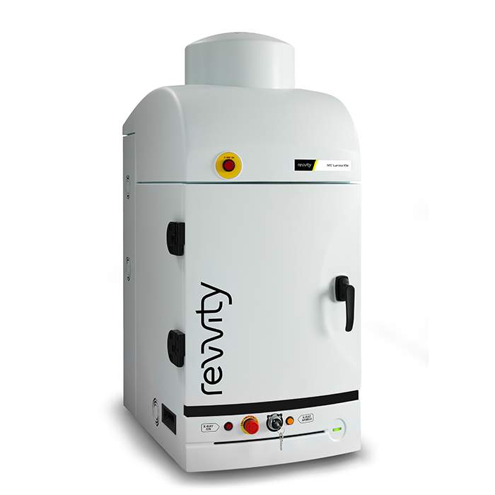
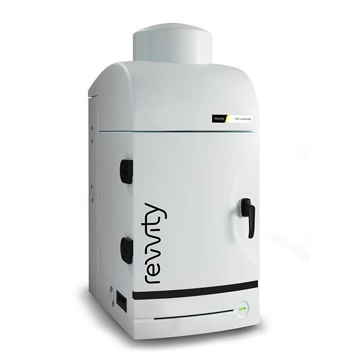
IVIS Lumina LT In Vivo Imaging System
- Bioluminescence
- Fluorescence
- Radioisotopic Cerenkov Imaging
- Compute Pure Spectrum Spectral Unmixing
- DyCE Imaging (Optional Upgrade)
- Extended NIR Range 150W Tungsten EKE
- Absolute Calibration to NIST® Standards
- Complimentary Living Image™ software licenses are provided with the IVIS systems and upon request.
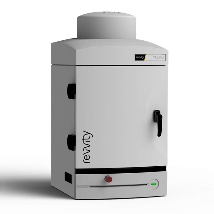
IVIS Lumina III In Vivo Imaging System
- Market trusted technology offering the fullest suite of leading imaging technologies, reagents and support
- Exquisite sensitivity in bioluminescence
- Full fluorescence tunability through the NIR spectrum
- Compute Pure Spectrum spectral umixing for ultimate fluorescence sensitivity
- Expandable system tailored to your workflow
- Complimentary Living Image™ software licenses are provided with the IVIS systems and upon request.
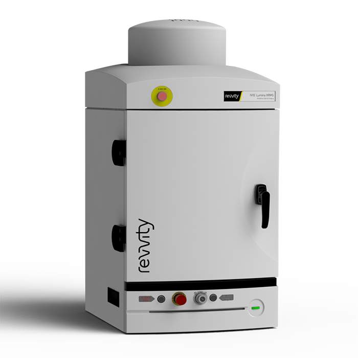
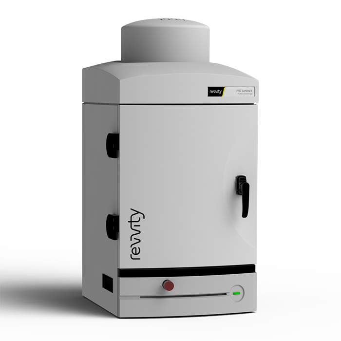
IVIS Lumina XRMS In Vivo Imaging System
- Multispecies optical and X-ray imaging
- Image mice, rats and other large animals
- High resolution, low dose digital X-Ray
- Exquisite sensitivity in bioluminescence
- Compute Pure Spectrum (CPS) spectral unmixing for ultimate fluorescence sensitivity
- Full fluorescence tunability through the NIR Spectrum
- Complimentary Living Image™ software licenses are provided with the IVIS systems and upon request.
IVIS Spectrum 2 series
- High sensitivity 2D and 3D bioluminescence & fluorescence imaging
- High throughput simultaneous imaging of up to 10 mice
- Fast data acquisition for rapid visualization of images in real-time
- Two powerful modes of fluorescence excitation - epi-fluorescence and trans-illumination
- Proprietary spectral unmixing algorithms for autofluorescence removal and multiplex imaging
- Easy, one-click co-registration with the Quantum GX3 microCT system
- Broadly adopted, easy to use, and intuitive, Living Image™ visualization and analysis software
- Integrated low-dose, ultra-fast microCT (IVIS SpectrumCT 2 only)
- Complimentary Living Image™ software licenses are provided with the IVIS systems and upon request.
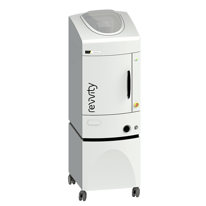
IVIS Spectrum 2 In Vivo Imaging System

IVIS SpectrumCT 2 In Vivo Imaging System
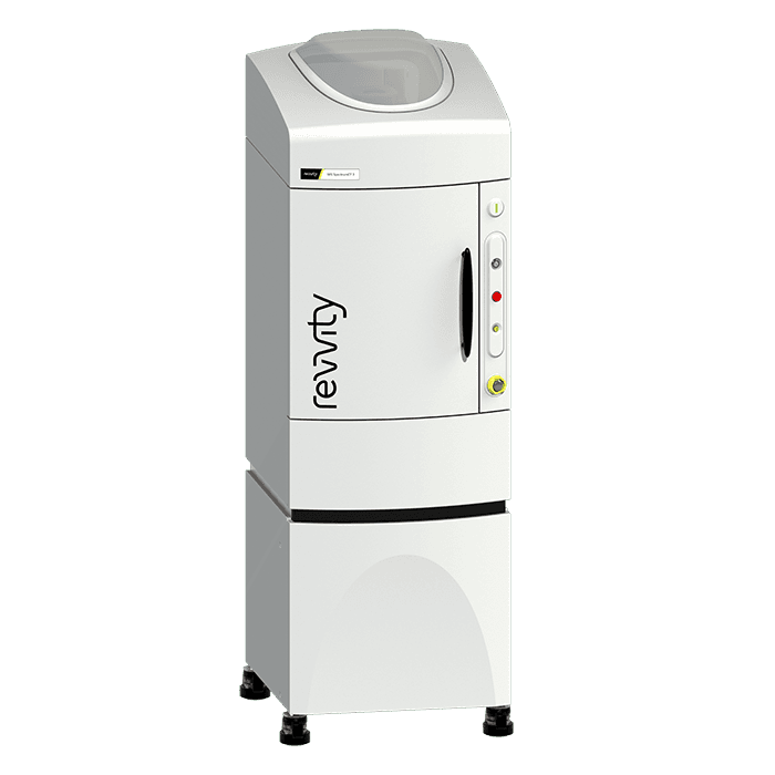
Quantum GX3 microCT imaging system
- Superior spatial resolution of 5 microns
- Wide FOV range from 8 mm to 86 mm
- Improved image-based respiratory gating
- Proprietary active ring reduction
- Continuous and step scanning modes
- Seamless co-registration with the IVIS 3D optical imaging system

Quantum GX3 microCT System
Featured resources


Filters
1 - 9 of 9 Products and Services

This product replaces the IVIS™ Spectrum system (part number 124262). As the leader in optical imaging with thousands of publications using our flagship IVIS Spectrum platform, the IVIS Spectrum 2 system is the next generation in optical imaging. Designed with an innovative camera with patented coating that delivers high sensitivity 2D and 3D bioluminescence and fluorescence imaging capabilities - the IVIS Spectrum 2 gives you the flexibility you need for your in vivo imaging studies.
This product replaces the IVIS™ SpectrumCT system (part number 128201). For ultimate ease and flexibility, the IVIS SpectrumCT 2 in vivo optical imaging system integrates 2D & 3D bioluminescence and fluorescence imaging with low-dose CT combining both molecular functional and anatomical in vivo imaging into single system.
The IVIS™ Lumina S5 high-throughput 2D optical imaging system combines high-sensitivity bioluminescence and fluorescence in a benchtop format. With an expanded 5 mouse field of view for 2D optical imaging plus our unique line of accessories to accelerate setup and labeling, it has never been easier or faster to get robust data- and answers- on anatomical and molecular aspects of disease.

The IVIS™ Lumina LT Series III pre-clinical in vivo imaging system offers the industries most sensitive benchtop platform at an entry level price.

High-Sensitivity Optical Meets High-Resolution X-rayThe IVIS™ Lumina X5 high-throughput 2D optical imaging system combines high-sensitivity bioluminescence and fluorescence with high-resolution x-ray into a compact system that fits on your benchtop. With an expanded 5 mouse field of view for 2D optical imaging plus our unique line of accessories to accelerate setup and labeling, it has never been easier or faster to get robust data- and answers- on anatomical and molecular aspects of disease.
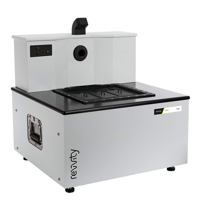
The Vega™ ultrasound system is Revvity's latest addition of leading preclinical in vivo imaging technology. With an innovative design, Vega is a hands-free automated ultrasound platform that delivers high-resolution 2D and 3D imaging in just a few minutes.Hands-free, automated, high-throughput ultrasound

Industry leading fluorescence, bioluminescence and x-ray imaging - All-In-One benchtop instrument! The IVIS™ Lumina XRMS (x-ray multi-species optical imaging system) adds to the versatility of the IVIS Lumina XR by offering the flexibility to image large animals up to 500 grams with optical and x-ray overlay.

The IVIS™ Lumina Series III brings together years of leading optical imaging technologies into one easy to use high sensitivity bench-top system.
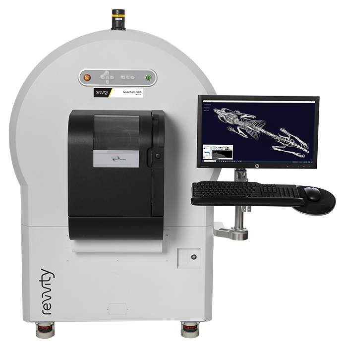
This product replaces the Quantum™ GX2 (part # CLS149276)The Quantum GX3 - advanced microCT system for ex vivo and in vivo imaging. The versatile Quantum GX3 microCT offers the flexibility researchers need by delivering the functionality to perform longitudinal studies, across a wide range of species, with fast, low-dose scanning and superior spatial resolution.